Ultrasound imaging, also referred to as sonography, is a diagnostic tool that utilises high-frequency sound waves to create images of the internal structures of the body. It offers a glimpse into the human body to assist healthcare professionals in diagnosing, monitoring, and sometimes even treating medical conditions, with a broad range of uses.
Ultrasound does utilise ionising radiation like other imaging options, therefore, the lower risks associated with ultrasound imaging has allowed it to become a diagnostic imaging modality of choice for diverse patient populations, including pregnant women.
What is an Ultrasound?
An ultrasound is a technique of diagnostic imaging that utilises high-frequency sound waves to produce images of tissues, organs and vessels within the body. A safe and non-invasive imaging technique, it facilitates assessment of foetal wellbeing during pregnancy as well without exposure to ionising radiation – making it a safe choice for all patients including pregnant women.
Ultrasound scans are safe and non-invasive, unlike X-rays which use ionising radiation and can introduce dangerous side effects on pregnancy. Ultrasounds only use a small transducer which sends out sound waves, the sound waves then reflect off organs, tissues and fluids within the body. The transducer then captures the echoes and a computer translates this information into images so that they can be viewed on a screen.
Accurate Ultrasound Scans, Fast Results
Private, convenient, and reliable—our London clinic ensures you get the answers you need quickly.

What sort of preparations do I need to do for an ultrasound scan?
Depending on the ultrasound you are having, the preparation for an ultrasound scan will vary. However, there are guidelines or instructions for preparation related to different types of ultrasounds, which can help make the experience easier and ensure the images taken are of the best quality.
- Wear loose, comfortable clothing to your appointment. You might also need changing into a hospital gown.
- For certain scans, particularly those of your abdomen, you may be asked to fast for a specified amount of time (typically 6 to 12 hours), prior to your appointment.
- In some specific pelvic ultrasounds, you may be asked to drink water and avoid urinating until your ultrasound has been completed, as a full bladder can enhance the visibility of your pelvic organs.
Some Common Types of Ultrasound
Ultrasound imaging is a cornerstone of diagnostics today, utilising high-frequency sound waves to facilitate imaging of the interior of the body. Important types of ultrasound include those tailored for the purpose of specific diagnostic evaluations:
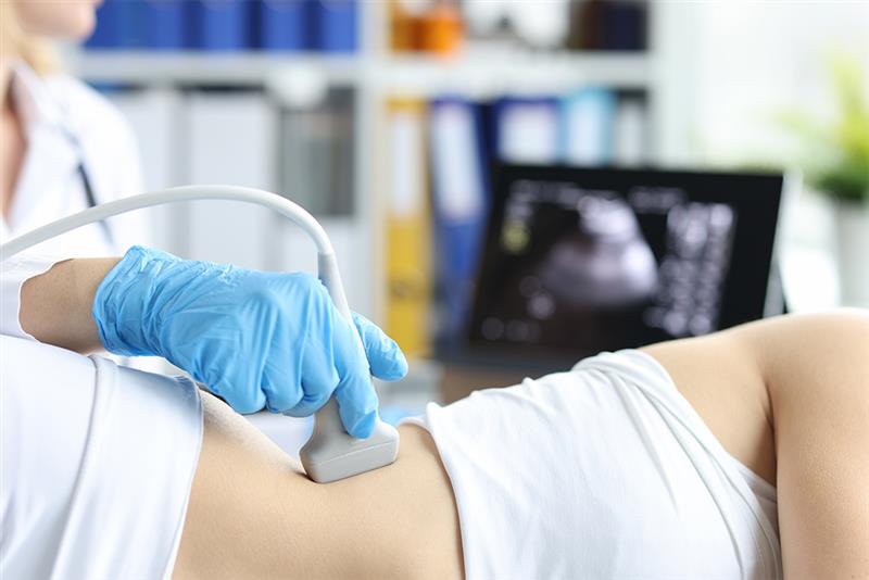
1. Abdominal Ultrasound
This kind of ultrasound is performed to visualise your abdominal organs, which may include your gallbladder, liver, pancreas, spleen and kidneys. It assists in the possible diagnosis of swelling, pain or infection in the abdomen.
2. Pelvic Ultrasound Imaging
Pelvic ultrasound involves a transvaginal or transabdominal method to assess pelvic organs, including the bladder, uterus and ovaries. Pelvic ultrasound is often used to find out the causes of abnormal bleeding, pelvic pain and other problems associated with menstruation.
3. Breast Ultrasound
Breast ultrasound evaluates breast tissues. It is often used with mammography to look for abnormalities observed on mammograms, including cysts and lumps, and to evaluate whether a mass is a fluid-filled cyst or a solid tumour.
4. Obstetric Ultrasound
These ultrasounds are conducted in pregnancy to monitor foetal development, examine foetal wellbeing, and establish the due date of delivery. It can also identify multiple pregnancies and increased foetal complications.
5. Transvaginal Ultrasound
This involves inserting the transducer into the vagina to obtain a closer view of the female reproductive organs – the ovaries, uterus and cervix. The transvaginal approach can provide clearer images for diagnosis.
6. Transrectal Ultrasound
These ultrasounds are used to assess the prostate gland and surrounding structures in males. This involves inserting the transducer into the rectum. Transrectal ultrasound would be used to look for symptoms such as difficulty urinating, or to direct procedures involving biopsy.
7. Carotid and Abdominal Aorta Ultrasound
This ultrasonographic test evaluates the carotid arteries in the neck that provide blood to the brain as well as the abdominal aorta. It is also useful in assessing for blood flow problems, blockages or aneurysms, which allows checking for risks of vascular disease or stroke.
8. Liver Ultrasound
This ultrasound is used to assess the liver for any abnormality, which may include tumours, fatty liver, or signs of liver diseases like hepatitis. It is also useful in determining liver size, shape and texture.
Accurate Ultrasound Scans, Fast Results
Quick appointments, expert staff, and detailed reports—all in a comfortable at Sono Clinic.
Uses of Ultrasound Techniques
Ultrasound techniques are used in all disciplines of medicine, due to their non-invasive nature, safety and ability to provide real-time images. Below is a review of how different ultrasound techniques are utilised in clinical practice:
1. Ultrasound for Baby Monitoring
This ultrasound is a non-invasive diagnostic method that may be used throughout pregnancy to assess the growth and development of the foetus. It can provide information regarding foetal growth (size and weight), placenta, and volume of amniotic fluid as needed, determine the foetus position, and detect congenital anomalies.
2. Breast ultrasound as a cancer detection method
This ultrasound is a diagnostic technique used to evaluate the breast for abnormalities, including breast cancer. It is mainly used with mammograms, particularly in women with dense breast tissue, where mammograms can be less effective.
3. Testicular ultrasound as a cancer detection method
Testicular ultrasound is an important diagnostic tool to evaluate testicular and scrotal abnormalities, including testicular cancer. It offers detailed images of the testicular and surrounding structures, which will assist your physician to look for cysts, masses or other abnormalities that may point to cancer or other conditions.
4. Ultrasound for abdominal pain
Ultrasound is routinely used to determine the cause of abdominal pain. An ultrasound will help to visualise the abdominal organs including the gallbladder, liver, pancreas, spleen and kidneys to look for problems such as kidney stones, gallstones, appendicitis and liver disease.
5. Ultrasound as it relates to muscles and joint pain
Ultrasound imaging is also utilised in the assessment and management of conditions related to tendons, muscles, joints and ligaments. Ultrasound can identify issues such as inflammation, sprains, tears or other soft-tissue injuries.
Specialised Ultrasound Techniques
To help solve specific diagnostic problems and to improve the applicability of ultrasound in a range of medical fields, specialised ultrasound techniques have been developed. Some of them that are now widely applied in clinical practice include:
-
Doppler Ultrasound
It is a specialised technique that assesses the blood flow moving through blood vessels, including the major veins and arteries of the neck, legs, and arms using the Doppler Effect. This helps assess speed and flow of blood to identify clots, blockages and constricted vessels.
-
3D and 4D Ultrasound
3D ultrasounds create three-dimensional images of the organs, tissues or foetus for more detail than a 2D ultrasound. 4D ultrasound allows for a dynamic 3D picture which shows real-time movement, including the movement of a foetus in uterus – Saline infusion sonohysterography (SIS).
-
Tubal Patency Ultrasound
Tubal Patency Ultrasound, also called hysterosalpingo-contrast sonography (HyCoSy), refers to a specialised ultrasound method developed to monitor tubal patency (openness) in women who are being evaluated for infertility.
The Importance of Ultrasound in Today’s Medicine
Ultrasound is incredibly valuable to today’s medicine, providing a non-invasive, versatile and affordable diagnostic tool that has reshaped how many disorders are diagnosed, treated, and managed. Here are some of the important points to convey the importance of ultrasound in today’s medicine:
-
Safety and Efficacy
Ultrasound is considered safe and effective in modern medicine. In contrast to other imaging modalities where ionising radiation (CT scan and X-ray) is present, ultrasound is a non-invasive procedure. This makes it a safer alternative for vulnerable populations as ionising radiation is unsafe for pregnant women and children. Therefore, there are very few cases in which an incision or injection is needed, thereby lowering the risk of complications and infection.
-
Application across a Variety of Disciplines
The greatest benefit of ultrasound technology is that it can be applied to numerous areas of medicine. This modality is not limited to use in obstetrics for monitoring foetal growth or stability, but is also used in radiology, emergency medicine, gastroenterology, cardiology and others. Ultrasound can create images of and organs, soft tissue blood flow – all with extreme clarity. This can help detect and manage numerous conditions during the lifespan of many patients.
Sono Clinic: Where Innovation Meets Ultrasound Excellence
Sono Clinic is recognised as one of the top diagnostic centres in the UK and specialises in diagnostic services for any medical condition. Our team carries out various diagnostic tests, including laboratory testing, imaging stud and radiology services.
Sono Clinic utilises advanced technology with experienced professionals, allowing for accurate and reliable results. Sono Clinic strives to offer comprehensive private ultrasound in London that helps for timely diagnosis and management of medical conditions. With a focus on quality and accuracy, we want to contribute to the overall health and wellbeing of our patients.