Pregnancy is a thrilling period in a woman’s life. However, it can also cause ambiguity and anxiety. One of the most vital considerations in your prenatal care is having routine ultrasounds to assess health and development of the foetus.
In this blog, we will address some frequently asked questions regarding ultrasounds while pregnant – why, when and more.
What is a pregnancy ultrasound?
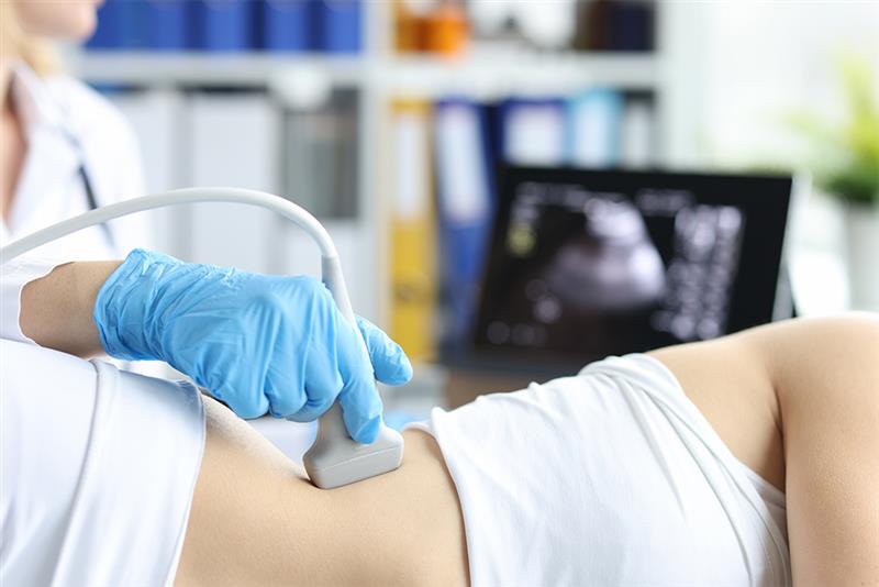
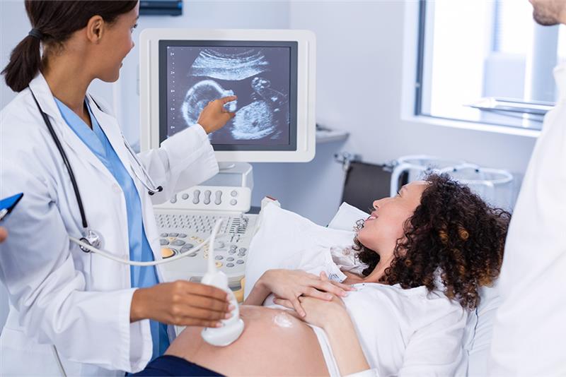
A pregnancy ultrasound, also called sonogram, is a process which uses sound waves to produce a picture of your growing baby in the uterus. It allows your doctor to:
- Evaluate the growth and development of the baby
- Assess the health of the placenta and uterus
- Look for any abnormalities or possible complications
Most women undergo one ultrasound around 18 to 20 weeks of pregnancy – in the second trimester. Some women may undergo an early ultrasound before 14 weeks – in the first trimester. This is done to confirm the pregnancy, provide an estimate for the due delivery date, and evaluate the growth and development of the baby in its early stage as well. The number of ultrasounds will vary among women as per their individual health conditions, such as history of obesity or asthma.
Fast & Reliable Ultrasound Scans in London
Skip the waiting lists and get a same-day private ultrasound with expert reporting.

Why is an Ultrasound necessary in Pregnancy?
Ultrasound is a non-invasive and simple test that gives vital information on your pregnancy and unborn baby to you and your doctor. During a pregnancy ultrasound, your doctor may:
- Confirm the viability of your pregnancy, meaning that everything looks normal and your baby’s heartbeat is going well.
- Identify whether you are having twins, triplets or multiples.
- Assess your baby’s age and also predict the due date for childbirth.
- Evaluate your baby’s movement, organs, muscle tone and development.
- Identify any pregnancy complications, such as ectopic pregnancy (a pregnancy growing outside of the uterus), a molar pregnancy (abnormal pregnancy tissue growth), or miscarriage.
- Identify the position of your baby’s head before birth.
- Evaluate the length of your cervix as well as the placenta position.
- Assess your baby’s health.
- Assess the size and growth of your baby.
- Examine your pelvic anatomy, including the cervix, ovaries and uterus.
Your healthcare provider may also use a pregnancy ultrasound for other testing and screening. Screening is a type of testing to see if your baby is more likely to have a certain condition compared to other babies. This type of testing is really important so the doctor can check for and manage any issues early on. Ultrasound can also be used for other pregnancy tests, like CVS (chorionic villus sampling) or amniocentesis, to check for genetic or chromosomal conditions like Edwards’ syndrome, Down’s syndrome and Patau’s syndrome.
Ultrasound is also an essential part of a biophysical profile (BPP), which is a test that uses ultrasound with a non-stress test. BPP testing observes whether a growing baby is getting enough oxygen and assists the doctor in recognising a possible concern and acting to keep your baby well. In short, ultrasound plays an important role in supporting your baby’s healthy development while allowing your doctor to make wise decisions about your ongoing pregnancy care.
Private Ultrasound Scans in London
Get fast, accurate, and confidential results—book your private ultrasound appointment today.
How Many Ultrasounds Will I Have During Pregnancy
The majority of pregnant women receive three to four ultrasound scans during pregnancy. The specific number of scans you receive, and the timing will depend upon your doctor’s recommendations and your underlying health issues. If you have a high-risk pregnancy or your doctor believes that there are issues affecting you or your baby’s health, additional ultrasounds may be recommended as a precautionary measure.
The following section explores different types of pregnancy ultrasounds and when they are typically performed:
6 to 14 Weeks (Dating Scan)
The ultrasound that you receive in the first trimester of pregnancy (6 to 14 weeks) is referred to as the dating scan. This pregnancy ultrasound can help your doctor to:
- Confirm the viability of your pregnancy
- Establish a due date for your child delivery
- Confirm that you have multiple pregnancies (more than one baby in your uterus)
- Confirm that your baby is developing in your uterus and you do not have an ectopic pregnancy
- Establish any issues that may create complications for you and your baby during the pregnancy
12 to 13 Weeks (Nuchal Translucency Scan)
The Nuchal Translucency Scan (NTS), often referred to as the 12-week scan, is typically conducted when you are 12 weeks pregnant. However, it can also be conducted during the 11th and 13th week of pregnancy. A scan during pregnancy examines the thickness of a small area at the back of your baby’s neck, known as the nuchal translucency. This helps assess the risk of your baby in having a genetic condition known as a chromosomal abnormality.
Similar to a dating scan, the nuchal translucency scan can monitor your baby’s growth and development, predict your due delivery date, establish the number of babies in uterus, and assess the presence of structural problems that may impact your baby’s health.
18 to 22 Weeks (Morphology Scan)
The scan conducted between 18 weeks and 22 weeks of pregnancy is known as the morphology scan or ‘foetal anomaly scan’. This specific ultrasound is intended for the following purposes:
- To assess the development of your baby’s internal organs
- To estimate your baby’s size and gestational age
- To monitor your baby’s heart rate and rhythm, and the location of your placenta in relation to your cervix
- To assess and measure your cervical length, ensuring that it is closed
32 to 36 Weeks (Third-trimester Ultrasound)
The ultrasound performed during the third trimester of pregnancy, usually between weeks 32 and 36, is called the third-trimester ultrasound. The doctor will use this ultrasound to:
- Evaluate the growth and position of the foetus and determine the feasibility of a normal delivery
- Assess the amount of amniotic fluid
- Check the position of the placenta
- Identify possible concerns, including congenital disabilities, low foetal weight or umbilical cord issues
This pregnancy ultrasound will help ensure a safe and healthy delivery for the mother and baby.
When Can You See a Baby on an Ultrasound?
Generally, you can see a baby on an ultrasound around six weeks of pregnancy. During this stage, you will likely see a gestational sac along with a small foetal pole on the scan. Generally, by seven to eight weeks, you will be able to see the heartbeat of your baby on the ultrasound.
What Ultrasound Is Most Important During Pregnancy?
All ultrasounds during pregnancy are essential. Every ultrasound assists the doctor in assessing the growth and development of your baby as well as your overall pregnancy health.
Are there any risks of ultrasounds?
Ultrasounds are largely regarded as safe during pregnancy and will not harm either the mother or the baby. However, physicians will suggest your ultrasounds are limited to prenatal checks authorised by them.
Book Your Private Ultrasound Scan in London
Receive quick results and professional care in a comfortable, discreet environment.
Conclusion
Ultrasounds during pregnancy are important to expectant mothers; they monitor the overall health and development of your baby, identify abnormalities and possible complications, and ensure timely intervention, if necessary.
If you want to know more about private ultrasound scans in London, have questions about your baby’s development, or are simply curious about this experience, speak to one of the healthcare professionals at our Sono Clinic. Our expert team of gynaecology and obstetric professionals offer great value and respect in providing top quality services to ensure you and your baby receive the necessary support and care for a healthy experience throughout your pregnancy journey.